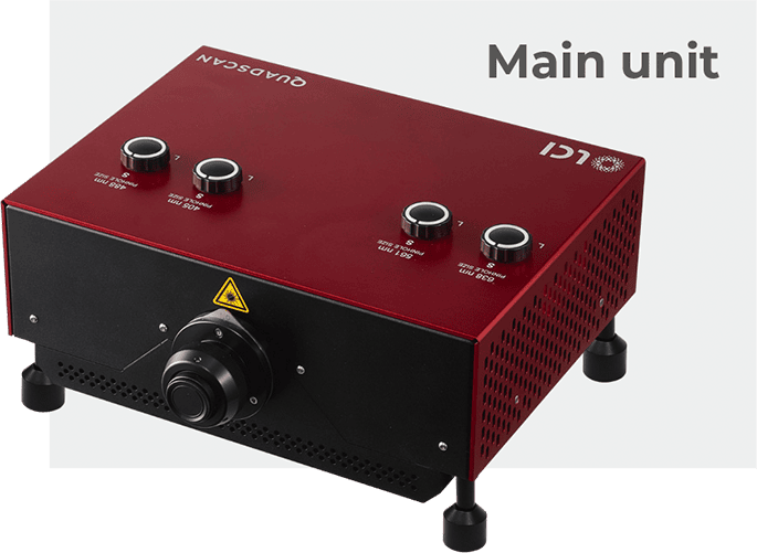
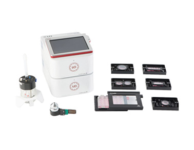
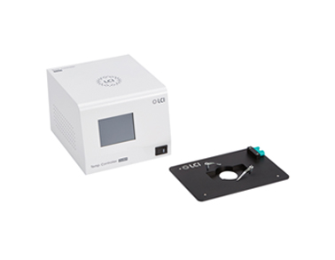
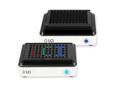
All-in-one structural module for each channel
QUADSCAN
- Overview
- Technology
- Imaging examples
- Microscope system
- Software
- Specifications
- Related Products
- Resources
QUADSCAN Built to do it all, Built for you
QUADSCAN is an advanced confocal imaging system that integrates a high-speed confocal unit, a motorized microscope, and powerful software into a single, streamlined platform.
Whether for live-cell imaging, time-lapse experiments, or high-resolution fluorescence analysis, QUADSCAN provide reliable and accurate biological results through advanced imaging technology, supporting high-quality experimental analysis.


Features
QUADSCAN has a unique subunit structure which contains all the necessary components for each fluorescence band excitation and detection in a single unit.
QUADSCAN supports single channel observation as well as up to four multi-channel (405 nm, 488 nm, 561 nm, and 638 nm) simultaneous excitation and observation
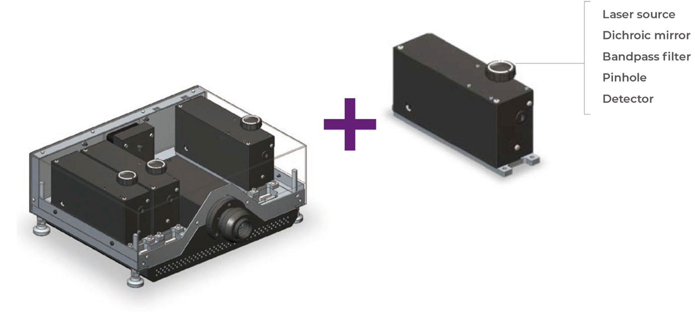
The subunit contains all the necessary componentsfor single band fluorescence excitation and detection, including a laser source,
a pinhole, and a detector.
a pinhole, and a detector.
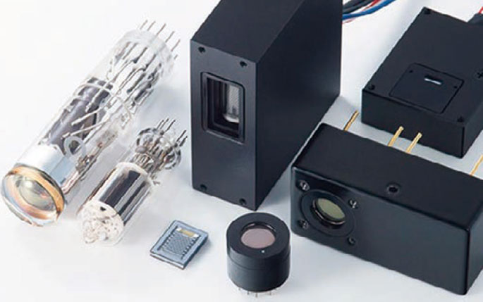
High-sensitivity detectors
By employing the world’s most sensitive detector (photomultiplier tube) and applying our signal processing know-how, it enables highly sensitive detection of fluorescence signals. This sensitivity is critical for enhancing signal-to-noise ratio in fluorescence imaging and expanding its applications across various biological fields.
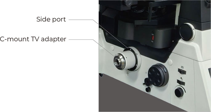
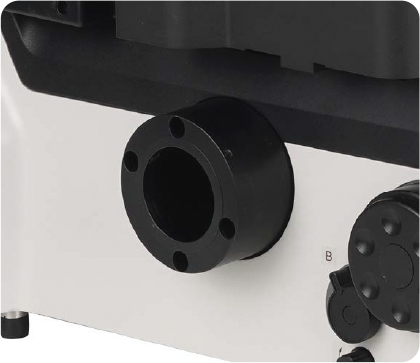
QUADSCAN is mounted via a side port of the inverted microscope
with a C-mount TV adapter.
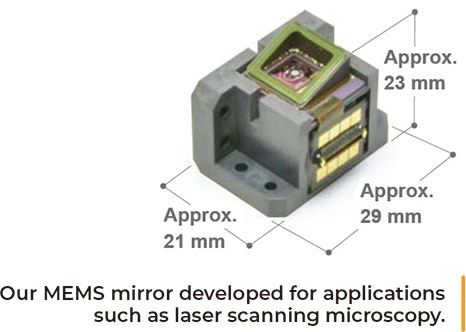
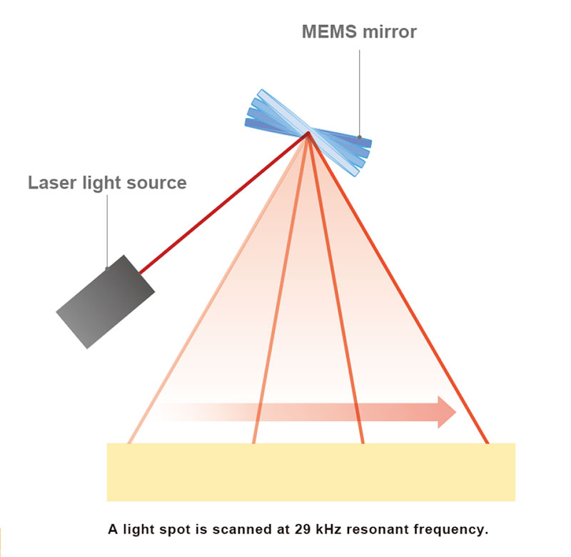
High-speed scanning with MEMS mirror
We adopted our 29 kHz resonant type high-speed MEMS* mirror as a spot scanning device. The MEMS mirror allows for a high-speed scan up to 76 frames/s and can be used for high-speed phenomena such as Ca dynamics.
The high-speed resonant scanning system reduces laser irradiation time, which enables low phototoxicity, low photobleaching and high-efficiency observation of live cells as well as fixed samples.
High-speed scanning allows for comfortable observation at high resolution with minimal display delay when searching for or focusing on samples.
* MEMS = Micro-Electro-Mechanical Systems
Conventional confocal optics
QUADSCAN was designed to comply to the conventional confocal optics, which has a long history, using a spot scanning device and pinholes to acquire optical sectioning images. When imaging thick fluorescence samples, only the fluorescence emitted from the focal plane of the objective passes through the pinholes to the detector, while fluorescence emitted from further away from the focal plane is blocked. It detects quantitative, reproducible and reliable signals. It also acquires high contrast images without the need for image processing techniques, such as deconvolution

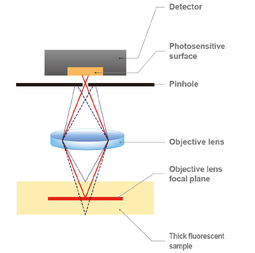
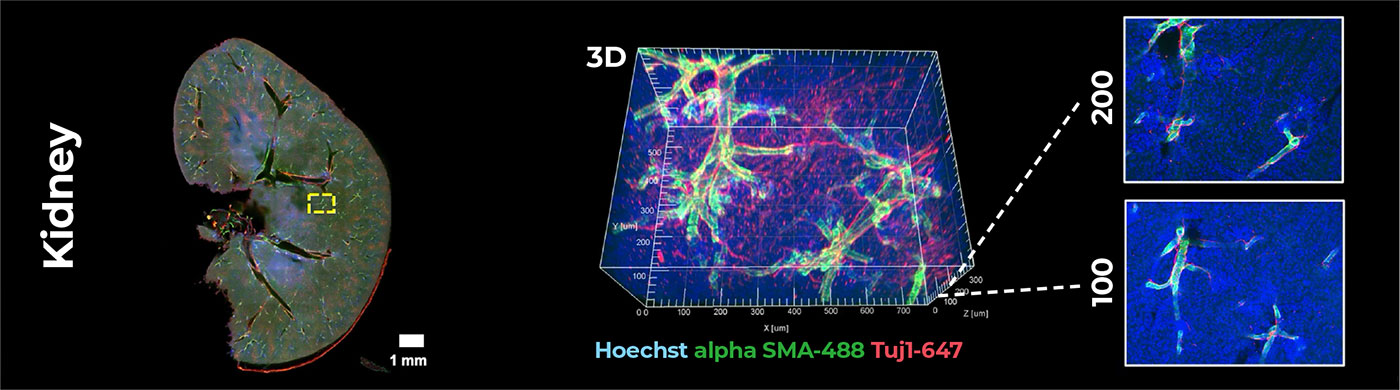
Sample: Mouse kidney section
QUADSCAN detects fluorescence in the objective focal plane only, so high contrast optical sectioning images can be acquired.
QUADSCAN detects fluorescence in the objective focal plane only, so high contrast optical sectioning images can be acquired.
Technology
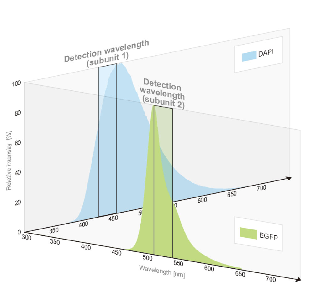
Enable simultaneous multicolor confocal acquisition without bleed-through
Conventional simultaneous multicolor observation
Fluorescent dyes emit a wide wavelength range of fluorescence, and if multiple fluorescent dyes are used, the fluorescence distributions are generally overlapped. When multiple dyes are excited simultaneously, an overlapping emission spectrum leaks into adjacent detection channels, causing an artifact of fluorescence imaging called bleed-through. The bleed-through cannot be prevented by simply using a dichroic mirror or a bandpass filter to select the wavelength, and this has been a problem
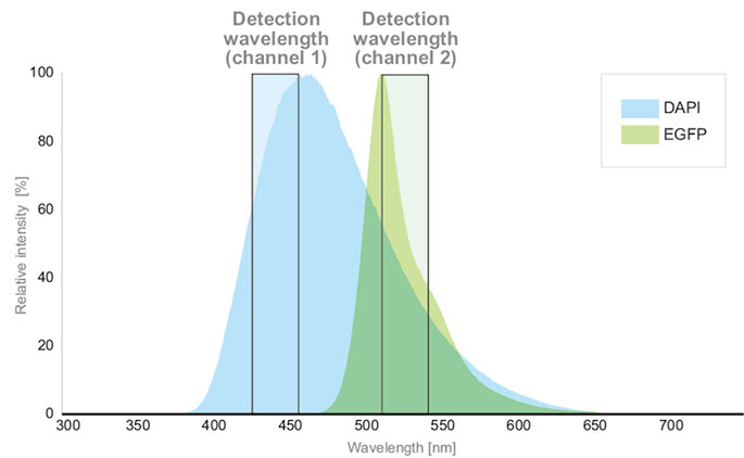
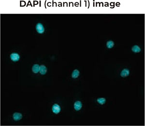
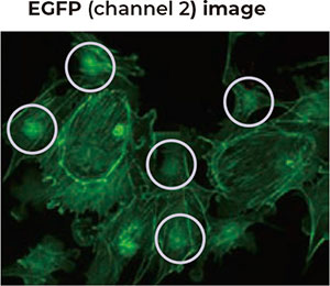
DAPI fluorescence leaks into the EGFP detection channel and causes artifact shapes of the nucleus in EGFP image. (Images are shown in pseudo-color.)
Newly developed
Simultaneous multicolor observation without bleed-through
Since QUADSCAN has a subunit structure, we can design the light path in each subunit independently. This minimizes the bleed-through to adjacent subunits and enables simultaneous multicolor observation with high detection efficiency.
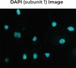
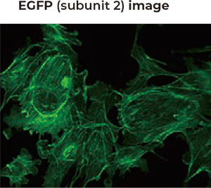
Simultaneous multicolor excitation does not cause DAPI fluorescence bleed-through
into the EGFP detection channel, and each fluorescent dye image is acquired accurately.
(Images are shown in pseudo-color.)
into the EGFP detection channel, and each fluorescent dye image is acquired accurately.
(Images are shown in pseudo-color.)
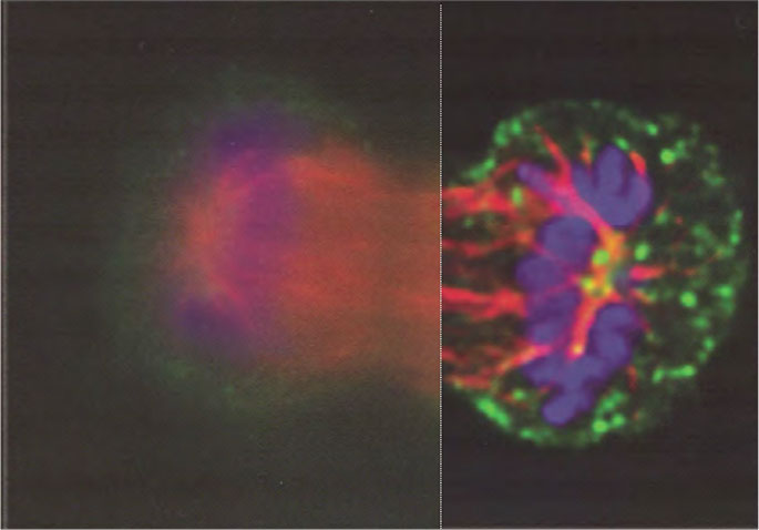
Imaging examples
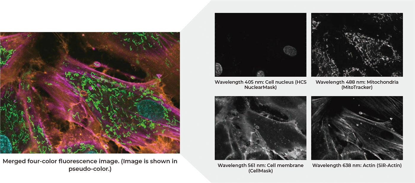
Live cell four-color imaging
Confocal imaging of cell nucleus, mitochondria, cell membrane, and actin filament with different dyes and channels. Each structure is clearly observed. 4-channel simultaneous imaging without frame drops
Sample: H9c2 cell line / Objective lens: 60x / Number of scan lines: 96
Laser wavelength: 405 nm, 488 nm, 561 nm, 638 nm
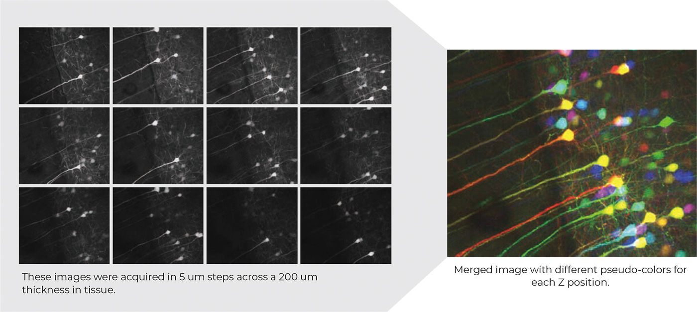
Z-section imaging of mouse brain
These optically sectioned images show that the structure of the cells and neurons in
this thick section of mouse brain are pyramidal in volumetric shape.
Sample and images courtesy of Dr. Christian Jüngst and Dr. Astrid Schauss (CECAD Imaging Facility, University of Cologne)
Sample: Fixed sample of transgenic mouse brain (Thy1-eYFP)
Objective lens: 20X (NA: 0.40)
Laser wavelength: 488
Application examples
- • Live cell imaging
- • High-speed Ca2+imaging
- • Membrane potential dye imaging
- • Time-lapse imaging
- • 3D/4D imaging
4D imaging
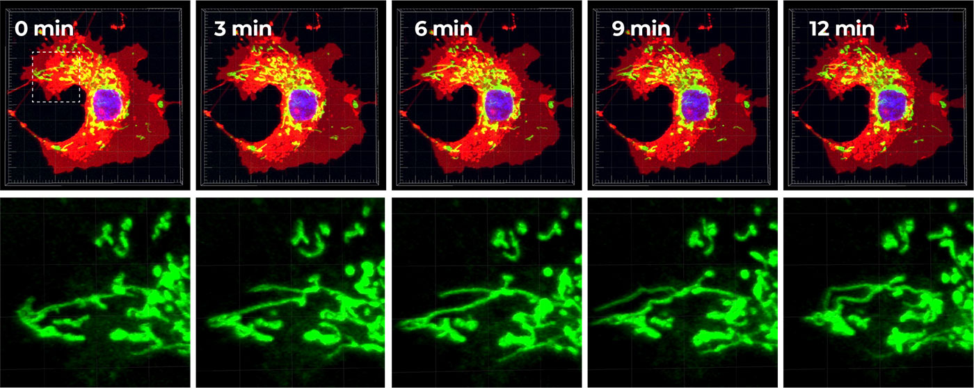
Sample: COS-7 Cell line / Dye: Hoechst (blue), Mito Tracker-488 (green), Cell Mask-deep red (red)
Lens: 60x (NA: 0.17) / Time-lapse: 3 min interval, 12 min total
3D Scale: 60 x 60 x 8 (μm) (z step interval: 1 μm)
Cell & Tissue Image
Cell multi-color imaging
Wavelength and detection sensitivity
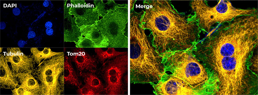
Sample: PFA fixed COS-7 Cell
Staining: DAPI (blue), Phalloidin-488 (green), Anti-Tubulin (yellow), Anti-Tom20 (red)
Lens: 60x (NA: 0.17)
Tissue multi-color imaging
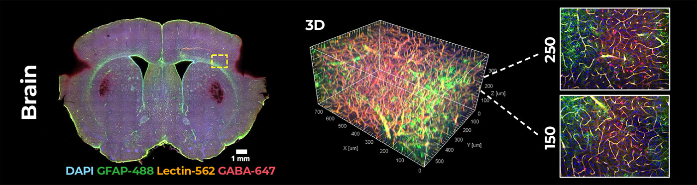
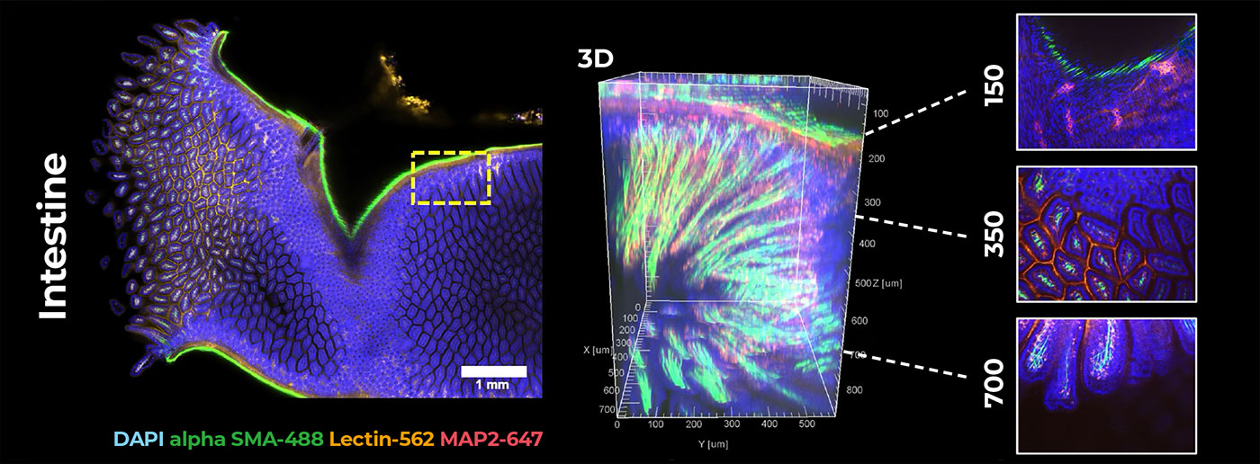
Sample: Various tissue slices (500 μm ~ 1 mm thickness) processed with optical clearing
Tissue Clearing Method: SCARF-based tissue clearing (crayon technologies )
Lens: 10x (NA: 0.4) / 3D scale: 550 x 630 x (400~850) μm (Z section: 3 μm )
| Type | Wavelength | Detector |
|---|---|---|
| Main unit | 405 nm | Standard type |
| 488 nm | High sensitivity type (Crystalline photocathode/ GaAsP) | |
| 561 nm | ||
| 638 nm |
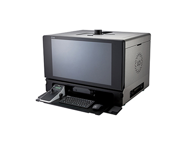
MIcroscope system

Stable and highly scalable live cell imaging platform
QUADSCAN provides an advanced imaging platform with 25mm field of view, to meet experimental needs of large flux observation. Fulll-motorized QUADSCAN realize integration of multimodal imaging technology. Anti-focus shift module can be optional which is more suitable for long-time dynamic imaging of living cells. QUADSCAN provides stable and reliable platform for confocal.
In addition, the touch screen could achieve smooth control of electric components while recording status of each sensor, recording user usage habits, switch application scene by one-click. Combined with powerful acquisition and analysis software, QUADSCAN is full of stability and scalability, meet a variety of experimental needs, provide reliable, repeatable data.
Incomparable 25mm field of view
With the research trend towards large-scale, high-flux and intelligent solutions, demand for faster data acquisition and higher throughput is increasing day by day. No matter it is bright field imaging or fluorescence imaging, QUADSCAN large-area illuminator and large-field fluorescence attachment provides uniform and bright cell imaging. Large size sensors and imaging interfaces that truly maximize performance of large format detector and provide perfect imaging platform for the future as camera technology continues to evolve rapidly.
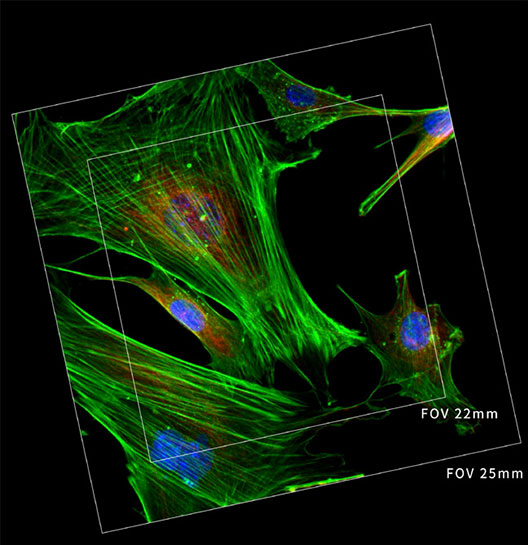
Large aperture observation optical system
Equipped with large diameter tube lens to expand light flow, cooperate with large target surface CMOS sensor, both bright field and fluorescence imaging could FOV25mm field of view.
FOV25 imaging objective
The objective with superior image flatness guarantees high quality images. Maximize potential of FOV25 objectives significantly speeds up data collection.
FOV25 imaging objective
The objective with superior image flatness guarantees high quality images. Maximize potential of FOV25 objectives significantly speeds up data collection.
Ideal living cells microscopic experiments platform
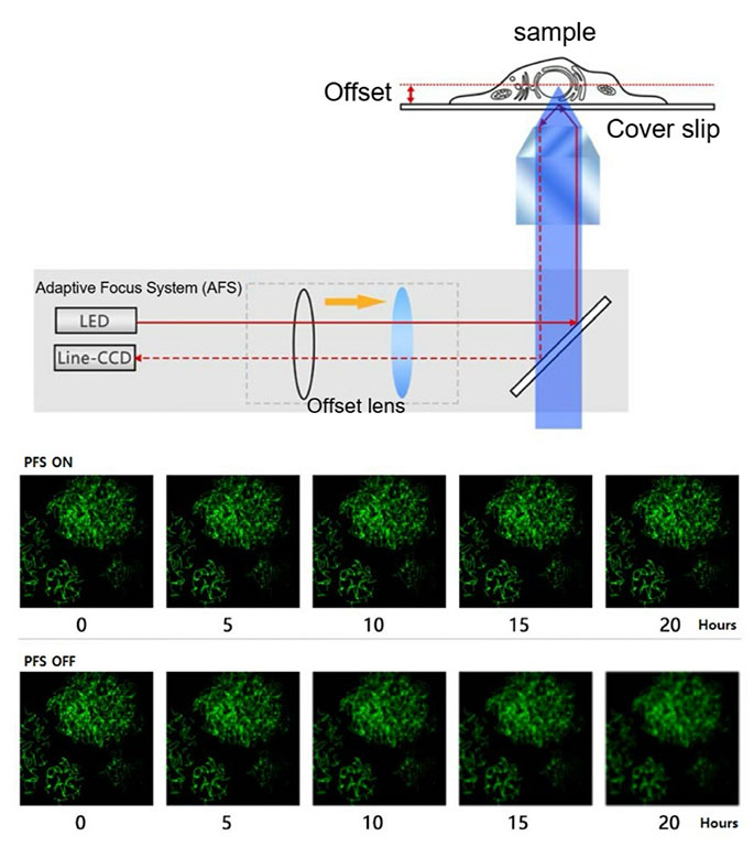
Even small changes of temperature and vibration in imaging environment can greatly affect stability of focus, adaptive anti-focus shift system (AFS) eliminates focus drift through static and dynamic measurements, highbrightness ED fluorescent light source and live cell culture system enables QUADSCAN realize multi-day living cell time-lapse observation, making it an ideal imaging tool for more demanding focused experiments.
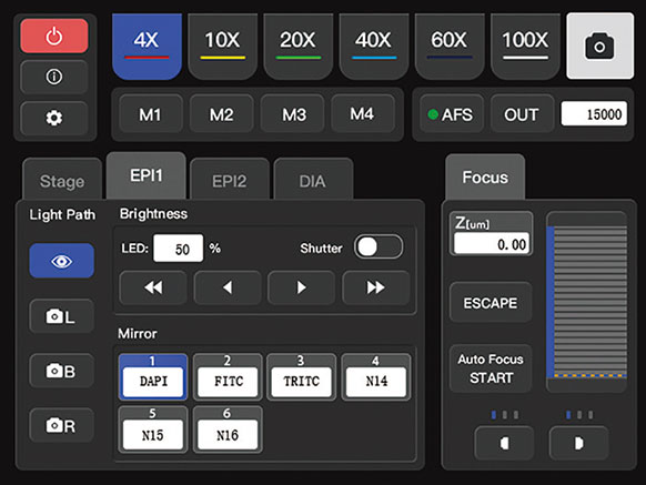
Front 5.6 inch touch screen
QUADSCAN was creatively designed front panel as a touch screen, which makes human-computer interaction more convenient, powerful and expandable. Left and right sides reserved knobs and buttons, even in a dark lab, it is easy to control, which allows researchers to focus more on the experiment itself, rather than complex microscope operation. fluorescence imaging could FOV25mm field of view.
AFS realize stable and reliable imaging results
Focusing stability is key factor of live cell imaging, even in constant temperature and humidity environment, focus position of microscope also varies slightly because of temperature and vibration changes, which greatly affects long-term observation of living cells. QUADSCAN adopt independent focusing design, maximumly reduce impact on Z axis from other mechanical components. Brand new designed AFS (Adaptive Focus System), adaptive anti-focus shift system, eliminate focus shift. Even when using high- magnification, highnumerical aperture objectives and advanced imaging technologies, such as super- resolution, confocal, TIRF, can still be clearly focused
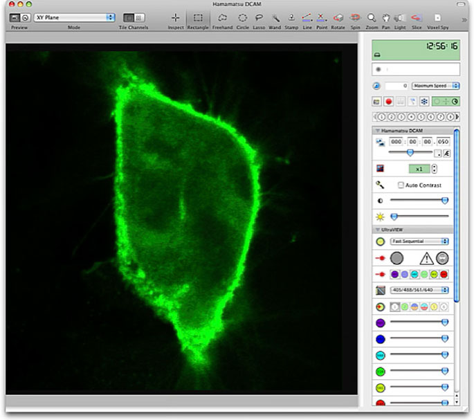
QuadVue
Acquisition
For faster 3D Image capture
QuadVue Acquisition is designed for high speed, 3D image capture. It provides a flexible capture interface to a wide range of cameras, microscopes and associated hardware, so that you can select the most suitable hardware items for your application without compromise. Capture protocols can be quickly constructed using the clear and easy to use Acquisition dialogue – from simple 2D image capture to multichannel 4D experiments. Flexible protocol design for all your imaging experiments. Tailor your experiment to the biology: QuadVue Acquisition is configurable with different hardware, and once set up in preferences you can construct complex and varied acquisition and analysis protocols without the need for complex macro writing skills.
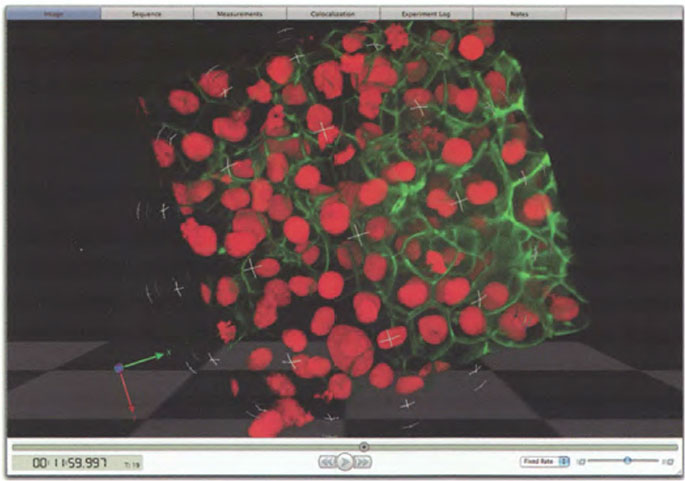
Visualization
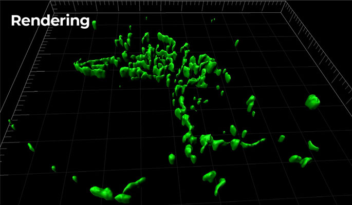
High resolution rendering of 3D and 4D volumes
QuadVue Visualization provides a choice of interactive renderers which allow you to view and interact with your images as 3D objects, in either fluorescence or isosurface mode, so that you can decide how to best display your data. In addition, QuadVue Visualization includes the Ray Tracer, a non-interactive renderer which produces very high quality rendered images, with user defined options such as lighting position, shadowing effects and background choices. The results can be spectacular in terms of the final image, although the process is more computationally intensive than the interactive rendering modes. This technique is ideal for producing publication quality images, when only the best will do.
Restoration
4D volume deconvolution
QuadVue Restoration provides a choice of proven restoration algorithms to improve the quality and resolution of 3D data sets quickly and easily. Applied to standard wide field fluorescence microscope images, the result is superior quality confocal data. Confocal, spinning disk and multi-photon images may reveal even more detail through the removal of out-of-focus haze. QuadVue Restoration is an essential imaging tool and supports a wide range of file formats from confocal microscopes and wide field systems.
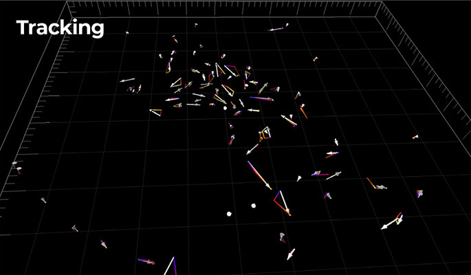
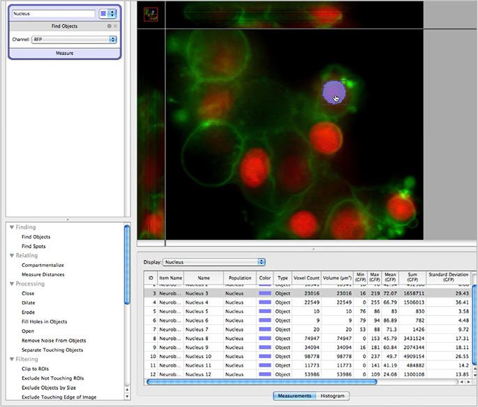
Quantitation
Identify, measure, track and analyze
QuadVue Quantitation provides a comprehensive range of options to analyze structure and function in 2D,
3D and 4D. Perform morphological analysis, measure fluorescence localization and colocalization and study trends. Present and publish images with measurement overlays, measurement tables, statistical data, charts and tracks. Analysis applications include FRAP, FRET, colocalization and ratiometric analysis.
Specifications
Confocal Unit
| Item | Specification | |
|---|---|---|
| Maximum effective field of view | 8.0 mm × 6.0 mm | |
| Maximum number of pixels | 1280 (H) × 960 (V) | |
| Image size & Frame rate (Typ.) | 1280 (H) × 960 (V): 19 frames/s 1280 (H) × 480 (V): 38 frames/s 1280 (H) × 240 (V): 76 frames/s |
|
| Zoom function | 1x, 2x | |
| Detection wavelength | at 405 nm excitation | 425 nm to 465 nm |
| at 488 nm excitation | 510 nm to 540 nm | |
| at 561 nm excitation | 580 nm to 619 nm | |
| at 638 nm excitation | 660 nm to 730 nm | |
| Detector | PMT, high-sensitivity GaAsP PMT | |
| Digital output | 12 bit | |
| Image acquisition mode | Single channel measurement, multiple channel sequential measurement (frame by frame), multiple channel simultaneous measurement (up to 4 channe | |
| Pinhole | 3 manual selections (large/medium/small) for each wavelength |
|
| Interface | USB 3.0 | |
| Output trigger connecto | SMA | |
| Lens mount | C-mount | |
| Power consumption | 90 VA | |
| Ambient operating temperature | +18°C to +28°C | |
| Ambient operating humidity | 30% to 80% (with no condensation | |
| Ambient storage temperature | -10°C to +5 °C | |
| Ambient storage humidity | 85% (with no condensation) | |
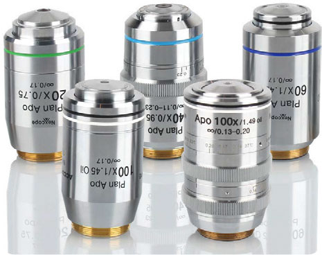
* Compatible objective lens : We recommend use of an objective lens with an image-side NA
(objective lens NA/magnification) smaller than 0.0375.
QuadVue Software
| Item | Specification |
|---|---|
| Acquisition | Full Agnostic Workflow, Multi dimensional acquisition, (X, Y, Z, time, wavelength, stitch) Stage Map, Dual cam |
| Visualization | 3D Visualization, Ray-Tracer, Surface Rendering |
| Quantitation(Optional) | Segmentation, 3D quantitation, Colocalization, Ratio Analysis, FRET/FREP Analysis |
| Restoration(Optional) | Deconvolution, Real-Time Deconvolution |
Inverted Microscope
| Item | Specification |
|---|---|
| Optical system | Infinite optical system |
| Eyepiece | 10×(22), with diopter adjustment -5 ~ +5 10×(25), with diopter adjustment -5 ~ +5 |
| Viewing tube | Seidentopf binocular observation tube, 10-40 degree tilt, pupil distance 47-78mm, eyepiece interface Ф 30 |
| Viewing tube base | eyepiece/camera (100/0, 0/100) |
| Relay lens | 0.5X, 0.7X, 1X |
| Objective | APO objective, S-APO phase contrast objective (optional) |
| Nosepiece | Motorized six-hole nosepiece(expansion slot), M25*0.75;AFS module included |
| Motorized stage | Electric control (grating type) : travel range 130mm x 100 mm (table 445 mm x 300 mm) Maximum speed: 25mm/s; Moving accuracy : 0.1μm Repetition accuracy: 0.5μm |
| Focusing system | Electric control drive, coarse fine tuning 3-gear switch, stroke: 8.5mm above the focus and 1.5mm below; Minimum step 0.01um |
| Touch LCD | In front of main body, display control light source intensity, objective magnification, fluorescence band, intermediate magnification, turntable position, bertrand lens etc.; |
| Intermediate magnification switching |
Manual 1 x, 1.5 x switching |
| Port | Electric switching spittingl ratio: left side: visual =100:0; right side: visual =100:0; bottom: visual =100:0; |
| Illumination system | Transmitted kohler illumination, 3W LED illumination |
| EPI illumination: white LED | |
| Condenser | 7-hole manual turntable, condition detection, condenser NA=0.52, WD=30mm |
| Fluorescent turntable | 6-hole electric fluorescent turntable (DAPI, FITC, mCherry standard); Ф 25/30 light pass; |
| Attachment (optional) | phase contrast, Hoffmann phase contrast, ND filter; DIC prism insert can be placed in nosepiece slot; DIC polarizer; analyzer slide plate; |
The product and software package names noted in this brochure are trademarks or registered trademarks of their respective manufactures. The products described in this brochure are designed to meet the written specifications, when used strictly in accordance with all instructions. The university, institute, or company name of the researchers, whose measurement data is published in this brochure, is subject to change.
The measurement examples in this brochure are not guaranteed. Specifications and external appearance are subject to change without notice.
Related Products
Resource
전체 6
|
Pamphlet QUADSCANQUADSCAN
|
2025.05.29 | 2025.05.29 |
|